Le tadalafil possède une affinité marquée pour la PDE5, mais épargne en grande partie les isoformes PDE1, PDE2 et PDE11, réduisant ainsi le risque d’effets extra-caverneux. L’action se traduit par une augmentation contrôlée de la circulation sanguine locale, indépendante des variations alimentaires. Sa pharmacocinétique repose sur une absorption digestive rapide, un métabolisme hépatique par CYP3A4 et une distribution tissulaire large. La biodisponibilité reste stable, et l’équilibre plasmatique est atteint en quelques jours lors d’administrations répétées. Les interactions cliniquement significatives surviennent avec les inhibiteurs puissants de CYP3A4 tels que le kétoconazole. Dans la littérature pharmacologique, acheter cialis 20 mg est souvent associé à des schémas d’utilisation basés sur la durée prolongée de son action.
Early perioperative death associated with reexpansion pulmonary edema during liver transplantation
Early Perioperative Death Associated With Reexpansion
Pulmonary Edema During Liver Transplantation
Wagner C. Marujo, Flavio Takaoka, Rita M. A. Moura, Fernando L. Pandullo,Andre R. Morrone, Marcelo M. Linhares, Alexandre Teruya, and Isaac AltikesHydrothorax is a frequent finding in patients with end-
REPE during a LT that rapidly led to the patient’s
stage liver disease. During the hepatectomy phase of liver
demise and speculate if this condition has not been
transplantation, it is often needed to evacuate large pleu- ral effusions. The acute expansion of the collapsed lung can cause reexpansion pulmonary edema with variable clinical significance. However, this complication has rarely been reported after liver transplantation. In conclu- Case Report sion, we report on an overwhelming reexpansion pulmo- nary edema during a liver transplantation that rapidly led
A 47-year-old male farmer with Child-Turcotte-Pugh C cir-
to the patient’s demise and speculate if this condition has
rhosis secondary to recurrent hepatitis B underwent hepatic
not been under recognized in the transplantation setting.
retransplantation. Preoperative cardiovascular and respiratory
(Liver Transpl 2005;11:1439-1443.)
work-up showed only moderate right pleural effusion.
After induction of the anesthesia, the ventilator was set to
conventional parameters (fraction of inspired oxygen: 40%;
Noncardiogenicacutepulmonaryedema(PE)may positiveend-expiratorypressure,5cmH O)and2large-bore
complicate the perioperative course of patients
central catheters were placed into the right internal jugular
undergoing liver transplantation (LT).1,2 Using radiog-
vein. Arterial blood gases and hemodynamic measurements
raphy and partial pressure of oxygen, arterial/fraction of
were within the normal range. Induction of immunosuppres-
inspired oxygen ratio Ͻ300 as diagnostic criteria,
sion consisted of 1 g bolus of methylprednisolone that was
Aduen et al. found PE in 52% of the patients undergo-
administered immediately before portal reperfusion. Cal-
ing LT.3 Immediate PE occurred in 25% of the
patients, 9% had late PE (developing de novo in the
During the operation, in the process of taking down dense
first 16 to 24 hours), and 18% had persistent PE (devel-
adhesions, the right diaphragm was inadvertently severed and
oping immediately and persisting for at least 16 hours).
the pleural space entered. Two and a half liters of icteric
Immediate PE had little clinical consequence, resolving
pleural effusion were rapidly drained out. We inserted a chesttube, but negative pressure was not applied. Table 1 depicts
within 24 hours, but persistent permeability-type PE
relevant respiratory and hemodynamic findings throughout
portended a worse outcome. This diffuse edema has
the procedure. No major hemodynamic changes occurred
been attributed to multiple etiologies: fluid overload,
after clamping the inferior vena cava and portal vein. Veno-
transfusion-related acute lung injury (TRALI), acute
venous bypass was not used. Immediately after portal revas-
respiratory distress syndrome (ARDS), and PE associ-
cularization, there was a short period (less than 2 minutes) of
ated with fulminant hepatic failure.2 The underlying
systemic hypotension (mean arterial pressure, 32 mm Hg),
pathophysiologic disturbances involve either an imbal-
accompanied by mild bradycardia (heart rate, 62 beats/min).
ance in the transcapillary hydrostatic forces (hydro-
Vasopressors were not used, and the patient recovered spon-
static-type PE) or a disruption of the permeability bar-
taneously. Only a few minutes following the revascularization
A rare cause of noncardiogenic PE is a form of a
Abbreviations: PE, pulmonary edema; LT, liver transplantation;
nondiffuse type of pulmonary endothelial injury that
TRALI, transfusion-related acute lung injury syndrome; ARDS,
occurs following rapid air expansion of a collapsed lung,
acute respiratory distress syndrome; REPE, reexpansion pulmonary
the so-called reexpansion pulmonary edema (REPE).4
In the LT setting, large pleural effusions are a common
From the Transplantation Program, Hospital Alema˜o Oswaldo
finding and are frequently evacuated during the proce-
Received March 9, 2005; accepted August 22, 2005.
dure. It is well recognized that the rapid evacuation of
Address reprint requests to: Wagner C. Marujo, MD, Liver Trans-
large volumes of air or fluid from the pleural space can
plantation Program, Hospital Alema˜o Oswaldo Cruz, Rua Prof. Arthur
cause REPE with variable clinical significance. How-
Ramos, 96, cj. 111; Sa˜o Paulo—SP, 01454-905, Brazil. Telephone: 55-11-
ever, this complication after LT was only reported by
3816.6655; FAX: 55-11-3816.7130; E-mail: wmarujo@uol.com.brCopyright 2005 by the American Association for the Study of
Jabber et al., who described a single case with a benign
Published online in Wiley InterScience (www.interscience.wiley.com).
Herein, we report an overwhelming perioperative
Liver Transplantation, Vol 11, No 11 (November), 2005: pp 1439 - 1443Table 1. Perioperative Variations of Hemodynamic and Ventilatory/Respiratory Parameters
Abbreviations: ICU, intensive care unit; HR, heart rate; MAP, mean arterial pressure; CVP, central venous pressure; PAWP, pulmonaryartery wedge pressure; MPAP, mean pulmonary artery pressure; CI, cardiac index; PVRI, pulmonary vascular resistance index; FiO2,fraction of inspired oxygen; PEEP, positive end-expiratory pressure; Sat O2, arterial saturation of oxygen. *Clinical onset of pulmonary edema. †Values between parentheses represent significant transient variations in the parameter during that period of time. ‡Evolution of the variables within that period of time.
of the portal vein, the oxygenation deteriorated (arterial oxy-
coagulopathy (international normalized ratio, 3.2), hypergly-
gen saturation, 85-90%) and a large volume of icteric plasma-
cemia (234 mg/dL), only mild metabolic acidosis (arterial
like fluid started pouring out from the endotracheal cannula.
blood – pH, 7.31; PCO , 38 torr; base excess, Ϫ6.4 mEq/L)
Pulmonary pressures and vascular resistance indexes increased
and normal serum potassium. The calculated partial pressure
20%, but left heart filling pressures remained within the nor-
of oxygen, arterial/fraction of inspired oxygen at the arrival at
mal range. Over the following 4 hours, an extra volume of
the intensive care unit was 236 (normal Ͼ300). The chest
3.0 L of the same fluid was suctioned from the endotracheal
tube was apparently functioning properly. Chest auscultation
cannula. Throughout the rest of the procedure, frequent but
revealed almost no breath sounds in the lower right chest and
short episodes of oxygen desaturation (arterial oxygen satura-
crackles in the mid portion of the same side. Auscultation on
tion, 80-86%) occurred, requiring transient increases of frac-
the left side was jugged to be normal. Electrocardiography was
tion of inspired oxygen (up to 60%) and vigorous endotra-
unremarkable. The mean pulmonary artery pressure and pul-
cheal suction. Positive end-expiratory pressure level remained
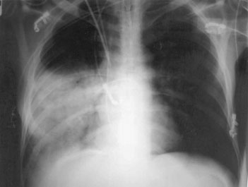
monary vascular resistance index increased. The chest radiog-
raphy (Fig. 1) showed a homogenous condensation in the
The operation was technically demanding but uneventful.
lower and middle lobes of the right lung. Intraoperative
Cold ischemia time was 19.5 hours. The anhepatic phase
lasted 58 minutes. Upon arterial reperfusion, the macroscopic
Over the following 5 hours, 2.7 L of a fluid similar to the
appearance of the liver was unremarkable. One hour later,
pleural and ascitic fluids were aspirated from the trachea. The
there were objective signs that the graft was working properly:
aspirate was slightly frothy, non – blood stained, and had total
production of good-looking bile, satisfactory urinary output,
protein and bilirubin ratios of 0.8, compared to plasma. Dur-
recovery of normothermia, and no overt clinical coagulopa-
ing the same period, the peritoneal and chest drains collected
thy. Throughout the procedure, 10 units of packed blood
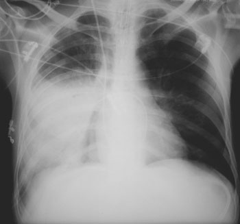
830 mL and 470 mL, respectively. A chest film taken 4.5
cells, 22 units of fresh frozen plasma, and 10 units of platelets
hours after the patient’s arrival at the intensive care unit
showed the same radiographic pattern depicted earlier (Fig.
Upon arrival at the intensive care unit, the patient was
2). Hemodynamic monitoring continued to show unremark-
awakening from anesthesia and needed to be sedated. The
able left heart filling pressures. We increased the positive
urinary output was adequate and the ascitic fluid was not
end-expiratory pressure level to 10 cm H O and placed the
frankly hemorrhagic. Despite relatively high serum levels of
patient in a slightly right lateral decubitus. We then managed
aminotransferase (aspartate aminotransferase, 3,400 IU/L;
to correct the patient’s fluid balance but we were not able to
alanine aminotransferase, 2,500 IU/L), other laboratory tests
improve his cardiac output using inotropic drugs. The pul-
suggested good allograft function: moderate laboratory
monary artery wedge pressure mildly increased (18 mm Hg)
but the cardiac index fell (2.7 L/min per m2). Despite aggres-sive ventilatory and hemodynamic support, severe hypoxemiaand a sustained increase in pulmonary vascular resistanceindex (872 dynes.s.cmϪ5) and mean pulmonary artery pres-sure (47 mm Hg) were followed by refractory hemodynamicinstability. The patient died 6 hours after arriving at the unit.
The autopsy determined the cause of death as bi-lobar
(lower and middle right lung lobes) reexpansion pulmonaryedema associated with circulatory collapse secondary to rightheart failure and sustained hypoxemia. In the affected pulmo-nary lobes, the alveolar spaces were filled with edema fluid. Large macrophages with the cytoplasm full of acidophilicfluid and scattered neutrophils and mononuclear cells werealso seen in the alveolar space. The pulmonary epithelium wasintact. The alveolar interstitium was moderately expanded byedema. There was a mild septal pulmonary capillaritis pro-duced by a mixed infiltration of neutrophils, macrophages,and mononuclear cells. The nonaffected lung depicted onlymild interstitial edema. The liver pathology showed mild tomoderate harvesting injury. Figure 2. Chest film taken 4.5 hours after the patient’s arrival at the intensive care unit showing the same radio- graphic pattern depicted earlier. Discussion
Noncardiogenic diffuse PE is a frequent finding imme-diately following LT and may significantly contribute
terfly wing” signal) is a mild enlargement of the heart
to the perioperative morbidity and mortality.1,2 A very
and major pulmonary vessels and a heterogeneous lung
common perioperative scenario is the occurrence of
infiltrate, especially in central and dependent areas, usu-
hydrostatic PE caused by water and sodium retention
ally accompanied by pleural effusions. However, iso-
and low oncotic pressure observed in end-stage liver
lated overhydration is particularly difficult to recog-
disease. This picture can be aggravated by the almost
nize.6 In any case, the interstitial PE begins to be
inevitable fluid overloading and overhydration associ-
mobilized in the first 24 to 72 hours, in a process that
ated with transfusion of multiple blood products, sig-
can be expedited with forced diuresis and adequate
nificant crystalloid infusion, and marked intraoperative
postoperative fluid management. However, in severe
fluid shifts. The typical radiographic appearance (“but-
cases, hypoxemia may delay the weaning process fromthe ventilatory support.
The occurrence of permeability-type, diffuse PE in
the LT setting is apparently less common. The inci-dence of clinically relevant events is less than 1%.2 Themost likely reason is TRALI. It has been postulated thatTRALI is induced by granulocytes that aggregate in thepulmonary microvasculature after activation by trans-fusion-derived antibodies, biologically active lipids, orother yet unidentified agents.7 Its true incidence isunknown and the attendant morbidity and mortalitymay be underappreciated. TRALI is characterized bythe rapid onset of respiratory distress, hypoxia, andnoncardiogenic PE during or soon after transfusion ofblood products. Most cases last only 2 to 6 hours. How-ever, mortality rates as high as 25% have been re-ported.8 Radiographic examination reveals diffuse and
Figure 1. Chest film immediately after the patient’s
fluffy infiltrates. Treatment is based only on aggressive
arrival at the intensive care unit showing condensation in
respiratory support.9 ARDS is another form of PE that
the lower and middle right lung lobes, signs of mild fluid overload and no significant pleural effusions. The chest
can occur following LT. Infection accounts for approx-
tube and central catheters were properly located.
imately half of the cases and its occurrence in the LT
setting has been considered an ominous sign. A diffuse
alveolar reexpansion, triggering endothelial activation
damage involves both the endothelial and epithelial
and neutrophil sequestration (priming event). This
surfaces and disrupts the lung barrier. Cardinal clinical
event would be followed by local enhancement of the
features of ARDS are the insidious onset of refractory
inflammatory cascade by attracting and activating cir-
hypoxemia and bilateral pulmonary infiltrates, but no
culating proinflammatory cytokines, antibodies and
primed cells generated upon reperfusion, hepatic fail-
REPE is a form of non-diffuse, permeability-type PE
ure, or TRALI, resulting in endothelial damage and
that occasionally occurs following acute evacuation of
capillary leak (second-hit).2 In addition, the release of
air or fluid from the pleural space, allowing rapid expan-
inflammatory mediators into the systemic circulation
sion of a long period collapsed lung. The underlying
that occurs following gut-liver reperfusion can cause
mechanism of REPE is not well defined. It is likely
significant systemic endothelial injury.14,15 Likewise, a
caused by an inflammatory response secondary to
severe harvesting injury might provide the milieu to the
expansion-related mechanical stress and reperfusion
release of inflammatory mediators into the systemic
injury to the alveolar-capillary membrana.11 Its inci-
circulation.14 Despite relatively good allograft function
dence varies from 14% in clinical observations, up to
in the immediate postoperative period, the presence of
27% in animal models.4 Following the precipitating
very early and high elevations of aminotransferase seems
event, the condition has been observed within the first
to support that the graft suffered a significant preserva-
hour in 64% of the patients or within 24 hours in the
tion injury. In this complex scenario, a significant har-
remainder.12,13 The process usually resolves in less than
vesting injury might have indirectly contributed to the
72 hours, but mortality rates as high as 20% have been
distinctive clinical course. Moreover, a systemic endo-
thelial injury, with a high incidence of pulmonary
During LT, evacuation of large volumes of pleural
edema, is frequently observed in patients with fulmi-
effusion is not rare, and REPE might have been under-
recognized. REPE may be a contributing factor for the
Despite the fact that REPE is 1 of the causes of PE
respiratory distress that frequently occurs in the early
and its main clinical manifestation (acute respiratory
postoperative period. Because of its ordinarily benign
distress) is indistinctive of other etiologies of PE, some
and transient clinical course, associated with nonspe-
clinical and radiological features clearly distinguish
cific radiographic signs, REPE findings may be over-
REPE from the other etiological entities, as follows: (1)
lapped by other perioperative pulmonary complications
absence of either clinical, hemodynamic or laboratorial
following LT, such as pleural effusions, atelectasis,
data to support cardiogenic PE etiology—normal/low
TRALI, and hydrostatic PE. REPE usually occurs in the
pulmonary artery wedge pressure; (2) REPE is not a
lung that was collapsed, but it can oddly happen in the
diffuse injury of the lung and is always related to rapid
evacuation of air or fluid from the ipsi- or contralateral
In the only other reported case of REPE during LT,
pleural space; (3) characteristic radiological features of
despite significant hypoxemia, the clinical course was
REPE are patchy or homogenous consolidation cir-
benign.5 In ordinary cases, less than 20 mL of frothy
cumscribed to segments or lobes of the affected lung;
sputum is usually eliminated over the first 30 minutes
(4) the characteristic radiological picture of TRALI or
of the acute phase. Up to our patient’s demise, an aston-
ARDS is a bilateral “white-out” infiltrate; and (5) REPE
ishing volume of almost 6 L flooded into his pulmonary
does not respond to forced diuresis and is not accom-
alveoli in 8 hours. Factors occasionally present in the
panied by elevated heart filling pressures.
LT setting, such as fluid overloading and TRALI, might
The diagnosis of REPE is based on clinical grounds,
have also contributed to this overwhelming flood.
indistinctive ipsi- or contralateral lobar consolidation
However, except for ARDS and its causative factors,
on radiography, and hemodynamic parameters incom-
diffuse-type PE usually resolves within 24-48 hours
patible with volume overload. The clinical features of
after LT and has little clinical consequences, irrespec-
REPE vary from asymptomatic radiographic findings
tive of etiology and physiopathogenesis. In fact, TRALI
alone to severe cardiopulmonary manifestation. Treat-
might be associated with a worse outcome.3
ment exclusively relies on ventilatory and hemody-
We speculate that a second-hit phenomenon might
namic support. Alternative forms of ventilatory sup-
explain the overwhelming magnitude of the pulmonary
port, such as high-frequency or independent lung
leak in this particular case. The alveolar-arterial mem-
ventilation may be able to provide better ventilation/
brane in the collapsed lung would be initially hit by
perfusion match.17 One should be aware that REPE
shear-stress and/or free-radical injury that occur upon
might occur in the rather complex and intricate LT
scenario with a dramatic clinical manifestation, eventu-
7. Silliman CC, Ambruso DR, Boshkov LK. Transfusion-related
ally leading to circulatory collapse.
acute lung injury. Blood 2005;105:2266-2273.
8. Toy P, Popovsky MA, Abraham E, Ambruso DR, Holness LG,
Kopko PM, et al. Transfusion-related acute lung injury: Defini-
Acknowledgement
tion and review. Crit Care Med 2005;33:721-726.
9. Sevransky JE, Levy MM, Marini JJ. Mechanical ventilation in
The authors thank the attendant surgeons of Pro-Figado for
sepsis-induced acute lung injury/acute respiratory distress syn-
their invaluable assistance in the operative procedure.
drome: An evidence-based review. Crit Care Med 2004;32(Suppl):S548 - S553.
10. Piantadosi CA, Schwartz DA. The acute respiratory distress syn-
References
drome. Ann Intern Med 2004;141:460-470.
11. Sherman SC. Reexpansion pulmonary edema: A case report and
1. Afessa B, Gay PC, Plevak DJ, Swensen SJ, Hemantkumar GP,
review of the current literature. J Emerg Med 2003;24:23-27.
Krowka MJ. Pulmonary complications of orthotopic liver trans-
12. Jackson RM, Veal CM, Alexander B, Brannen AL, Fulmer JD.
plantation. Mayo Clin Proc 1993;68:427-434.
Re-expansion pulmonary edema. Am Rev Respir Dis 1988;137:
2. Yost CS, Matthay MA, Gropper MA. Etiology of acute pulmo-
nary edema during liver transplantation: A series of cases with
13. Mahfood S, Hix WR, Aaron BL, Blaes P, Watson DC. Re-ex-
analysis of the edema fluid. Chest 2001;119:219-223.
pansion pulmonary edema. Ann Thorac Surg 1988;45:340-345.
3. Aduen JF, Stapelfeldt WH, Johnson MM, Jolles HI, Grinton SF,
14. Matuschak GM, Henry KA, Johanns CA, Lechner AJ. Liver-
Divertie GD, Burger CD. Clinical relevance of time of onset,
lung interaction following Escherichia coli bacteremic sepsis and
duration, and type of pulmonary edema after liver transplanta-
secondary hepatic ischemia/reperfusion injury. Am J Respir Crit
4. Matsuura Y, Nomimura T, Murakami H, Matsushima T, Kake-
15. Olthoff KM. Molecular pathways of regeneration and repair
hashi M, Kajihara H. Clinical analysis of reexpansion pulmonary
after liver transplantation. World J Surg 2002;26:831-837.
16. Trewby PN, Warren R, Contini S, Crosbie WA, Wilkinson SP,
5. Jaber S, Perrigault P-F, Souche B, Pouzeratte Y. Re-expansion
Laws JW, Williams R. Incidence and pathophysiology of pulmo-
pulmonary edema with normal pulmonary artery occlusion pres-
nary edema in fulminant hepatic failure. Gastroenterology 1978;
sure during liver transplantation. Intensive Care Med 2002;28:
17. Cinella G, Dambrosio M, Brienza N, Ranieri VM. Reexpansion
6. Groeneveld AB, Polderman KH. Acute lung injury, overhydra-
pulmonary edema with acute hypovolemia. Intensive Care Med
tion or both? Crit Care 2005;9:136-137.
Noruega Renova Contrato com Gemalto para Passaporte Eletrônico Biométrico AMSTERDÃ -- A Gemalto (Euronext NL0000400653 GTO), líder mundial em segurança digital, renovou o contrato plurianual para personalizar e emitir Passaportes Eletrônicos para os cidadãos noruegueses. A empresa conduz todo o processo de fornecimento, desde a produção do documento de viagem até a personalização e
Prevalence and management of rheumatoid arthritis in thegeneral population of Greece—the ESORDIG studyA. Andrianakos1,2, P. Trontzas3, F. Christoyannis1, E. Kaskani4, Z. Nikolia5,E. Tavaniotou1, A. Georgountzos3 and P. Krachtis1 for the ESORDIG study groupyObjective. To assess the prevalence and management of rheumatoid arthritis (RA) in the general adult population of Greece. Methods. This cr
 but the cardiac index fell (2.7 L/min per m2). Despite aggres-sive ventilatory and hemodynamic support, severe hypoxemiaand a sustained increase in pulmonary vascular resistanceindex (872 dynes.s.cmϪ5) and mean pulmonary artery pres-sure (47 mm Hg) were followed by refractory hemodynamicinstability. The patient died 6 hours after arriving at the unit.