Le tadalafil possède une affinité marquée pour la PDE5, mais épargne en grande partie les isoformes PDE1, PDE2 et PDE11, réduisant ainsi le risque d’effets extra-caverneux. L’action se traduit par une augmentation contrôlée de la circulation sanguine locale, indépendante des variations alimentaires. Sa pharmacocinétique repose sur une absorption digestive rapide, un métabolisme hépatique par CYP3A4 et une distribution tissulaire large. La biodisponibilité reste stable, et l’équilibre plasmatique est atteint en quelques jours lors d’administrations répétées. Les interactions cliniquement significatives surviennent avec les inhibiteurs puissants de CYP3A4 tels que le kétoconazole. Dans la littérature pharmacologique, acheter cialis 20 mg est souvent associé à des schémas d’utilisation basés sur la durée prolongée de son action.
Pii: s0090-4295(97)00700-0
ADULT UROLOGY
USE OF INTRALESIONAL VERAPAMIL TO DISSOLVE PEYRONIE’S
DISEASE PLAQUE: A LONG-TERM SINGLE-BLIND STUDY
JAMIL REHMAN, ALEXANDRU BENET, AND ARNOLD MELMAN
ABSTRACT Objectives. Multiple conservative therapies for the treatment of Peyronie’s disease have been offered with variable and poor response rates. Calcium channel blockers have been shown in vitro and in vivo to inhibit secretion and synthesis of extracellular matrix, including collagen, glycosaminoglycans, and fibronectin, as well as causing increased collagenase and anti transforming growth factor-beta activity. Calcium antagonists, including verapamil, are effective in stimulating the remodeling and degradation of extracellular matrix in tissue by altering the metabolic pathways of fibroblasts. Recently, a pilot study (1994) showed preliminary promising results in treating plaque caused by Peyronie’s disease. This randomized single-blind placebo- based study (1994 to 1996) was undertaken to confirm the hypothesis. Methods. In this randomized single-blind study, 14 patients completed the study and were divided into two groups: the verapamil treatment group (n ϭ 7) or the control saline group (n ϭ 7). Verapamil or saline was
injected directly into the Peyronie’s plaque once a week for 6 months. Patients were evaluated before and after treatment with duplex ultrasound to confirm the extent of the lesion and to measure volume of the plaque, and by interview and mailed questionnaire 3 months after treatment. Patients being treated with oral calcium antagonists were excluded from the study. Results. A decreased plaque volume was measured in 57% of the verapamil-treated men versus 28% in the control group (P Ͻ0.04). Penile curvature demonstrated an improvement trend of 37.71 Ϯ 9.3° to 29.57 Ϯ
7.3° in the verapamil-treated patients, but the difference was not significant (P Ͻ0.07). Plaque softening was
noted in all patients treated with verapamil. There was significant objective improvement in plaque-associated penile narrowing in all patients in the verapamil group. Subjective plaque-associated erectiledysfunction (quality of erection) showed improvement in 42.87% of the verapamil group versus none in thecontrol group (P Ͻ0.02). There was no local or systemic toxicity except for an occasional ecchymosis/bruise
at the injection site. After a positive clinical response, plaque size, penile angulation, and symptoms continued to improve. Decrease in plaque size was noted in each of the responders in the first 3 months. Conclusions. This randomized single-blind study suggests that intralesional injection of calcium channel blocker may be a reasonable approach in some selected patients for the treatment of Peyronie’s disease with noncalcified plaque and penile angulation of less than 30°. Patients whose plaque failed to respond to intralesional verapamil therapy within 3 months or whose angulation was greater than 30° at presentation were more likely to benefit from surgery. UROLOGY 51: 620–626, 1998. 1998, Elsevier Science Inc. All rights reserved. FrancoisGigotdelaPeyronie1,2iscreditedwith albuginea.Theplaquetypicallyformsonthedorsal
first describing a disease that is characterized
or dorsolateral surface of the penis under the neu-
by a fibrous plaque formation involving the tunica
rovascular bundle, but may involve any area of thecorpus cavernosum. Induration of the tunica albu-ginea results in focal loss of elasticity with im-
This article was presented (podium session) at the American Uro-
paired shaft elongation during penile erection. In
logical Association annual meeting, 1996.From the Department of Urology, Albert Einstein College of
the early stage of the disease, bending around the
Medicine/Montefiore Medical Center, Bronx, New York
plaque during penile erection may result in pain. In
Reprint requests: Arnold Melman, M.D., Department of Urol-
severe cases, the bend may interfere with vaginal
ogy, Albert Einstein College of Medicine, Montefiore Medical
penetration or may cause dyspareunia. In severe
Center, 210th Street, Bronx, NY 10467Submitted: July 14, 1997, accepted (with revisions): October
Peyronie’s disease, the penis distal to plaque (hour-
glass deformity) may be flaccid during erection. 620 ALL RIGHTS RESERVED
Although the exact etiology remains unknown,3,4
was 16 months (range 11 to 24). The patients were evaluated
the following theories have been proposed: fre-
by detailed history, physical examination, biothesiometry, and
quent chronic irritation,1 vasculitis,5 genetic caus-
penile plethysmography. Plaque length was measured by cal-ipers (Vmoeleov OP-270, Germany) and duplex ultrasound to
es,6–10 delaminate injury,11 inherited predisposi-
confirm the extent of the lesion and to measure volume of the
tion and repeated trauma,12 autoimmune insult,13
plaque. The volume of the plaques averaged 1.4 cc (range 1.5
to 2.7) before therapy. Seven patients had been treated unsuc-
Despite numerous options for treatment, none
cessfully with oral vitamin E, and 1 patient had new plaque
have been clearly successful and the disease re-
formation after previous Nesbit plication. Twelve patientscompleted the treatment in 6 months and 2 patients com-
mains a therapeutic dilemma for urologists.15 Var-
pleted treatment in 4 months (total n ϭ 14). Of the 2 patients
ious oral drugs including vitamin E,16,17 para-ami-
who completed the study in 4 months, 1 left the state because
nobenzoate (POTABA),18 procarbazine,19–22 col-
of family emergency and the other dropped out because of
chicine,23 tamoxifen,24 intralesional injection of
personal reasons. Both of these patients were available for final
steroids,24,25 alone or in combination with hyal-
interview and examination. Patients being treated with oralcalcium antagonists were excluded from the study. The pa-
uronidase, collagenase,26,27 orgotein,28–32 parathy-
tients were divided into treatment group (n ϭ 7) and control
roid hormone,33 dimethyl-sulfoxide (DMSO),29 in-
group (n ϭ 7). The patients in the treatment group received
terferon,34 a combination of para-aminobenzoate
intralesional injections of verapamil in an isotonic saline ve-
and vitamin E,35 ultrasound therapy along with hy-
hicle, whereas the control group received injections of saline
drocortisone therapy,36 and vitamin E combined
alone. This study was approved by the institution reviewboard of the Albert Einstein College of Medicine/Montefiore
with hydrocortisone14 have been attempted. With
Medical Center, Bronx, NY. Inclusion criteria were as follows:
POTABA and colchicine there is a significant inci-
(1) age range 35 to 70 years with clinical evidence of Pey-
dence of gastrointestinal toxicity. In addition,
ronie’s disease, that is, pain and plaque along with deformity of
there is the danger of bone marrow depression
the penis of at least 1-year duration; (2) discontinuation of any
with colchicine. A controlled trial of POTABA in
previous oral or other medication for Peyronie’s disease for atleast 3 months. Exclusion criteria were any history of calcium
comparison to vitamin E failed to show any thera-
channel blocker therapy or therapy interfering with calcium
peutic benefit.20 Parathyroid injections have had
no long-term success.33 DMSO has been shown tocause cataract formation. Interferon-2 treated
TECHNIQUE
cultures of fibroblast from Peyronie’s disease ex-
After local penile blockage with 2% lidocaine, the verapamil
hibit inhibited fibroblast proliferation and collagen
or saline was injected into the plaque with a 10-mL syringe
production but there are no in vivo study reports.
and a 25-gauge needle once a week (Figure 1). Approximately
Calcium channel blockers have been shown in
4 to 8 plaque punctures were performed at each sitting in aneffort to distribute the drug uniformly through the plaque. The
both in vitro and in vivo studies to inhibit synthe-
verapamil was injected 1 mg/0.1 cc (10 mg/1 cc). The dosage
sis/secretion of extracellular matrix molecules, in-
ranged from 10 to 27 mg. This dosage was chosen after con-
cluding collagen, glycosaminoglycans, fibronectin,
sultation with a pharmacist, taking into consideration the re-
as well as increasing collagenase and transforming
sponse rate in the pilot study by Levine et al.37 The patient was
growth factor-beta (TGF-) activity. Calcium
asked to compress the injection site for 5 minutes to decreaseecchymosis. Blood pressure and heart rate were continuously
channel blockers such as verapamil are effective in
monitored throughout the procedure and after injection for
stimulating the remodeling and degradation of ex-
the first 3 months, and because no patient exhibited any drop
tracellular matrix in tissue. Because the concentra-
in blood pressure or any related cardiac effects, monitoring
tion of verapamil required to induce the degrada-
was discontinued for the remaining 3 months. No systemic,
tive metabolic response in fibroblasts in the
local, acute, or chronic toxicity was noted except for an occa-sional transient ecchymosis/bruise at the injection site. To
laboratory exceeds the maximum safe serum level
prevent the incidental injury to the dorsal nerve fibers or dor-
by 100-fold, only intralesional therapy is possible.
sal arteries, the needle was inserted into the dorsolateral or
At present, verapamil is the only calcium channel
lateral side depending upon the location of plaque. Precaution
blocker available in injectable form. A pilot study37
was taken not to instill the drug into the corpus cavernosum.
showed promising results with an intralesional in-
Slight gentle pressure into the syringe was required for injec-tion into the tunica albuginea, whereas instillation into corpus
jection of verapamil into plaques caused by Pey-
cavernosum did not require pressure.
ronie’s disease. The purpose of the present studywas to test the effect of intralesional verapamil in asingle-blind, placebo-based study. DATA ANALYSIS AND STATISTICS
Objective and subjective data were gathered at completion
of the study. Objective assessment was done by direct inter-
MATERIAL AND METHODS
view, physical examination, and measurement of plaquelength and width by two independent investigators with cali-
PATIENT SAMPLE
pers, confirmed by penile ultrasound. Subjective data were
A total of 18 men ranging in age from 37 to 67 years (mean
gathered by questionnaire 3 months after completion of the
52) entered into this randomized single-blind study. Four
study. The data are expressed as mean and standard error of
men dropped out in the first month of the study, 1 preferring
the mean (SEM). The groups were compared using the un-
surgical intervention and 3 because of scheduling conflicts.
paired Student’s t test at a significance of P Ͻ0.05 by statistical
The mean duration of the disease in these men prior to therapy
software (STATVIEW 4.5, Abacus Concepts, Berkeley, Calif).
UROLOGY 51 (4), 1998
to 6.3 Ϯ 0.44 post-treatment in the control group(P Ͻ0.02).
Most urologists interested in sexual dysfunction
in men agree that acute or repeated mechanicaltrauma (sexual or nonsexual) to the tunica albug-inea is delivered via the septal fibers, which areresponsible for maintaining the axial rigidity dur-
Cross section of shaft of penis and dorsal
ing erection. Trauma with an erect penis results in
neurovascular bundle. Part of the figure has been en-
tissue disruption and microvascular injury (focal
larged to show the relationship of the neurovascular
vasculitis) that leads to exudation of fibrin.5 Fibrin
bundle to the plaque and the injection technique. First
triggers fibroblast activation and proliferation that
local block is obtained with 0.2% lidocaine. Then the
results in excessive deposition of extracellular col-
verapamil (1 mg/0.1 cc [or 10 mg/1 cc] of plaque vol-
lagen matrix in the tunica albuginea.11,38,39 The
ume as determined by ultrasound or calculated as pre-
formation of excessive fibrous tissue may also be
viously mentioned) is injected into the plaque with a10-mL syringe and 25-gauge needle. Injections are
triggered in genetically predisposed individuals,
given weekly. Approximately 4 to 6 plaque punctures
either from abnormal fibroblastic activity or de-
are needed per injection to distribute the drug uniformly
creased fibrinolysis. Calcium channel blockers
through the plaque. The injection site should be com-
have been shown to inhibit synthesis/secretion of
pressed for 5 minutes to decrease ecchymosis. To pre-
extracellular matrix molecules, including collagen,
vent the incidental injury to the dorsal nerve fibers or
glycosaminoglycans, and fibronectin, as well as in-
dorsal arteries, the needle is inserted into the dorsolat-
creasing collagenase and TGF- activity. During
eral or lateral side depending upon the location of
the last two decades, tremendous achievements
plaque. Precaution is taken not to instill the drug deep
have been made in calcium channel blockers and
into corpus cavernosum. Slight gentle pressure into sy-
their role in collagen synthesis by fibroblast, both
ringe is always needed to inject into the tunica albug-
at the structural and biochemical levels. Better un-
inea as compared to easy instillation into the corpuscavernosum.
derstanding ultimately led to their clinical studiesin humans for fibromatosis. Calcium channelblockers such as verapamil are effective in stimu-
lating the remodeling and degradation of extracel-
The results are described in Tables I and II. All
lular matrix in tissue. Because the concentration of
patients who responded to verapamil did so in the
verapamil required to induce the degradative re-
sponse in fibroblasts in the laboratory exceeds the
(1) Plaque size and volume: plaque length de-
safe serum level by 100-fold or more, only intrale-
creased from 3.1 Ϯ 0.5 cm pretreatment to 1.8 Ϯ
sional therapy is possible at present.
0.4 cm post-treatment in the verapamil group ver-sus 2.9 Ϯ 0.5 cm pretreatment to 3.0 Ϯ 0.2 cm
FIBROBLAST, COLLAGEN METABOLISM, AND CALCIUM
post-treatment in the control group (P Ͻ0.03). CHANNEL BLOCKERS
Plaque width decreased from 1.7 Ϯ 0.95 cm pre-
Diegelmann and Peterkofsky40 provided evi-
treatment to 0.8 Ϯ 0.1 cm post-treatment in the
dence that calcium channel blockers alter cell
verapamil group versus 1.59 Ϯ 0.31 cm pretreat-
shape and tissue remodeling via epigenetic control
ment to 1.81 Ϯ 0.57 cm post-treatment in the con-
of the extracellular matrix. Ehrlich et al.41 de-
trol group (P Ͻ0.05). Plaque volume decreased
scribed experiments with antitubulin agents that
from 1.421 Ϯ 0.23 cm3 pretreatment to 0.63 Ϯ
resulted in inhibition of collagen synthesis and se-
0.19 cm3 post-treatment in the verapamil group
cretion of osteoblasts and fibroblasts. Dietrich and
versus 1.37 Ϯ 0.27 cm3 pretreatment to 1.39 Ϯ
Duffield42 demonstrated that calcium antagonist
0.29 cm3 post-treatment (P Ͻ0.04).
verapamil can alter synthesis of collagen and non-
(2) Penile curvature: penile curvature showed an
collagen proteins. Aggeler et al.43 demonstrated
improvement trend of 37.71 Ϯ 9.3° to 29.57 Ϯ 7.3°
that calcium antagonists as well as a calmodulin
in 28.57% of the verapamil-treated patients versus
blocker change fibroblast shape. They induced de-
no improvement in the control group, but the dif-
tachment of rabbit synovia fibroblasts from their
ference was not statistically significant (P Ͻ0.07).
substrate, resulting in altered morphology of the
(3) Quality of erections (subjective): the quality
fibroblast, decreased collagen synthesis, and a
of erections improved from 5.69 Ϯ 6.5 pretreat-
marked increase (20-fold) in the secretion of col-
ment to 7.4 Ϯ 0.37 post-treatment in 42.86% of the
lagenase. The altered cells were noted to alter the
verapamil group versus 6.28 Ϯ 0.52 pretreatment
secretion of extracellular matrix proteins on a con-
UROLOGY 51 (4), 1998 TABLE I. Response to intralesional verapamil injection Verapamil Verapamil Control Group Control Group Pretreatment Post-treatment Pretreatment Post-treatment Method of Parameter Measurement (Mean ؎ SEM) (Mean ؎ SEM) (Mean ؎ SEM) (Mean ؎ SEM) KEY: SEM ϭ standard error of the mean. * P Ͻ0.03 (plaque length post-treatment verapamil versus control group).
† P Ͻ0.05 (plaque width post-treatment verapamil versus control group).
‡ P Ͻ0.04 (plaque volume post-treatment verapamil versus control group).
§ P Ͻ0.07 (penile curvature post-treatment verapamil versus control group).
Ե Quality of erections scaled 1 to 10 (1 ϭ no erection; 10 ϭ very rigid erection). ** P Ͻ0.02 (quality of erections post-treatment verapamil versus control group).
centration-related basis, which correlates posi-
Kappas et al.49 found that rats with a surgically
tively with the degree of cell shape change. Once
created abdominal wound treated with verapamil
triggered, the increased rate of collagenase synthe-
(1 mg/kg administered intravenously) produced
sis persists for up to 1 week, even after the agents
significantly fewer peritoneal adhesions than con-
are removed and the cells reanchored onto the sub-
trols. Johnson et al.50 reported that nifedipine re-
strate. Kelly44 demonstrated that the secretion of
sulted in improved wound healing (skin and facial
extracellular matrix is calcium dependent (regulat-
wounds as well as enteric anastomoses) in animals
IN VITRO CELLULAR STUDIES AND CALCIUM CHANNEL CLINICAL HUMAN STUDIES, INCLUDING THE PRESENT BLOCKERS STUDY AND OTHER CALCIUM CHANNEL BLOCKER
In vitro experiments by Askey et al.45 found that
STUDIES
verapamil specifically inhibits fibroblast secretion.
Lee et al.51 demonstrated that intralesional injec-
Lee and Ping46 reported experiments with bovine
tion of verapamil hydrochloride directly into a
fibroblasts exposed to increasing concentrations of
burn scar markedly reduced its size. In a pilot, sin-
verapamil and nifedipine, and found decreased in-
gle arm study, Levine et al.37 used verapamil to
corporation of radiolabeled proline and sulfate into
dissolve Peyronie’s plaque and reported promising
extracellular matrix collagen and glycosaminogly-
results. The findings of the present randomized
cans, respectively. They demonstrated that treat-
single-blind study suggest that verapamil injection
ment of the fibroblast-populated collagen matrix
results in a significant decrease in plaque size. A
with calcium channel blockers (verapamil hydro-
decreased plaque length of 30% or more was noted
chloride [100 mol/L] and nifedipine [10 mol/L])
in 57.14% of patients treated with verapamil versus
markedly reduced the incorporation of tritiated
only a 28.57% decreased plaque length in the con-
proline into the extracellular matrices of the fibro-
trol group (P Ͻ0.04). There was no significant
blast-populated collagen matrix. Therefore, it ap-
change in penile angulation after treatment, al-
pears necessary to deliver verapamil directly to the
though a trend was noticed in one quarter
fibrous tissue to avoid high blood level. Fitscha et
(28.57%) of patients. The persistence of curvature
al.47 demonstrated, in a vascular lesion induced in
may mean that although plaque size is reduced, the
an animal model, that treatment with calcium
remaining collagen tissue is sufficient to cause
channel blocker (isradipine) treated fibroblasts re-
chordee. The more dramatic improvement in pe-
sults in decreased incorporation of proline and sul-
nile narrowing (bottleneck deformity) than curva-
fate into collagen and glycosaminoglycans, respec-
ture associated with plaque injection may be due to
the relatively thinner lateral extension of theplaque compared to the thicker midline plaque
IN VIVO ANIMAL STUDIES AND CALCIUM CHANNEL
with septal extension, as previously described.37
BLOCKERS
Penile pain improved in both groups though there
Steinleitner et al.48 have shown that verapamil
is a significantly more rapid improvement in penile
markedly reduced adnexal adhesions in rabbits.
pain in the verapamil group (mean 4 weeks) than
UROLOGY 51 (4), 1998 TABLE II. Response of patients to self-administered questionnaire regarding intralesional therapy Verapamil Group Control Group Parameter Post-treatment (n ؍ 7) Post-treatment (n ؍ 7) * Pain improved in both groups but more quickly in the verapamil group (4 weeks) than in the control group (10 weeks). † Excellent ϭ normal sexual function, residual deformity less than 15%.
‡ Moderate ϭ some improvement of erection or deformity 15°–30°, coitus possible.
§ Poor ϭ absent erections or deformity greater than 30°, coitus impossible.
in the placebo group (mean 10 weeks). There was
appears to induce rapid, beneficial results in some
also no worsening of sexual dysfunction during the
patients for reduction of plaque size (Figure 2).
study, which is significant because of the potential
The beneficial effects of intralesional verapamil are
injury to the dorsal neurovascular bundle of the
apparent within the first 3 months. For patients
penis. It seems calcium antagonists are best used
who respond to treatment, the injections should be
for immature plaque (plaque of less than 2-year
continued for 6 months. The patient who fails to
duration) with penile angulation of less than 30°.
respond to intralesional verapamil or whose angu-
On the other hand, histologic evaluation of ad-
lation is more than 30° at presentation should be
vanced disease has revealed islands of active fibro-
considered a candidate for surgery. Also, use of
blasts randomly dispersed throughout plaques. In-
calcium antagonists more potent than verapamil
tralesional injection of verapamil at multiple sites
should be considered as they become commer-
into plaque appears to allow verapamil to inhibit
cially available in injection form. The injection of
the activity of these fibroblasts in older plaque,
verapamil is clinically safe for patients with Pey-
which may not be possible by other methods.
ronie’s disease if precautions are taken to preventinjury to the dorsal neurovascular bundle. When
CONCLUSIONS AND FUTURE STUDY
compared with other methods of treatment, injec-
The injection of verapamil appears to be clini-
tion of verapamil appears to induce a rapid, bene-
cally safe for treating patients with Peyronie’s dis-
ficial effect in some patients (those with angulation
ease if used judiciously. When compared with
of less than 30°) for reduction of plaque size. Pa-
other methods of treatment, injection of verapamil
tients with localized plaque are the best candidates
UROLOGY 51 (4), 1998
Factors associated in the aetiology of Peyronie’s disease. Br J Urol 54: 748 –750, 1982.
4. Kaufman JJ: Peyronie’s: its cause. Scand J Urol Nephrol
138 (suppl): 219, 1991.
5. Smith BH: Peyronie’s disease. Am J Clin Pathol 45:
6. Nyberg LM Jr, Bias WB, Hochberg MC, and Walsh PC:
Identification of an inherited form of Peyronie’s disease with autosomal dominant inheritance and association with Du- puytren’s contracture and histocompatibility B7 cross-react- ing antigens. J Urol 128: 48 –51, 1982.
7. Willscher MK, Cwazka WF, and Novicki DE: The asso-
ciation of histocompatibility antigens of the B7 cross-reacting group with Peyronie’s disease. J Urol 122: 34 –35, 1979.
8. Somers KD, Winters BA, Dawson DM, Leffell MS,
Wright GL Jr, Devine CJ Jr, Gilbert DA, and Horton CE: Chro- mosome abnormalities in Peyronie’s disease. J Urol 137: 672– 675, 1987.
9. Bias WB, Nyberg LM Jr, Hochberg MC, and Walsh PC:
Peyronie’s disease: a newly recognized autosomal-dominant trait. Am J Med Genet 12: 227–235, 1982.
10. Guerneri S, Stioui S, Mantovani F, Austoni E, and Si-
moni G: Multiple clonal chromosome abnormalities in Pey- ronie’s disease. Cancer Genet Cytogenet 52: 181–185, 1991.
11. Devine CJ Jr, Somers KD, and Ladaga LE: Peyronie’s
disease: pathophysiology. Prog Clin Biol Res 370: 355–358, 1991.
12. Hinman F Jr: Etiologic factors in Peyronie’s disease.
Urol Int 35: 407– 413, 1980.
13. Rompel R, Weidner W, and Mueller-Eckhardt G: HLA
Scheme for treatment of Peyronie’s disease
association of idiopathic Peyronie’s disease: an indication of
plaque with intralesional verapamil or surgical interven-
autoimmune phenomena in etiopathogenesis? Tissue Anti-
tion. *Effect of size on responsive plaque to intrale-
gens 38: 104 –106, 1991. sional verapamil: plaque length less than 2.5 cm, 50 to
14. Novak GI, Burdina GV, and Salomatina LA: [Activity of
free radical processes in Peyronie’s disease]. Lab Delo 11: 42– 90% reduction in size; plaque length greater than 2.5cm, less than 50% reduction in 6 months.
15. Ludwig G: Evaluation of conservative therapeutic ap-
proaches to Peyronie’s disease (fibrotic induration of the pe- nis). Urol Int 47: 236 –239, 1991.
for intralesional injection of verapamil. For pa-
16. Scardino PL, and Scott W: The use of tocopherols in the
tients who respond to intralesional therapy and
treatment of Peyronie’s disease. Ann NY Acad Sci 52: 390 –
whose plaque size is less than 2.5 cm, the plaque
17. Scott WW, and Scardino PL: A new concept in the treat-
size may be reduced by 50% to 90% in 6 months,
ment of Peyronie disease. South Med J 41: 173–177, 1948.
but if the plaque size is greater than 2.5 cm, then
18. Hasche-Klunder R: Treatment of Peyronie’s disease
plaque will be reduced by less than 50% in 6
with para-aminobenzoacidic potassium (POTOBA). Urologe
months. It seems that the best use of calcium an-
[A] 17: 224 –227, 1978.
tagonists is for noncalcified plaque having a dura-
19. Oosterlinck W, and Renders G: Treatment of Peyronie’s
disease with procarbazine. Br J Urol 47: 219 –220, 1975.
tion of less than 2 years and with penile angulation
20. Morgan RJ, and Pryor JP: Procarbazine (Natulan) in the
less than 30°. Histologic evaluation of advanced
treatment of Peyronie’s disease. Br J Urol 50: 111–113, 1978.
disease has revealed islands of active fibroblasts
21. Aboulker P, and Benassayag E: Treatment of plastic
that are randomly dispersed throughout plaques. It
induration of corpora cavernosa penis with procarbazine.
is conceivable that intralesional injection of vera-
J Urol Nephrol 76: 499 –503, 1970.
pamil at multiple sites into such plaque may allow
22. Bystrom J: Induration penis plastica. Experience of
treatment with procarbazine Natulan. Scand J Urol Nephrol
verapamil to inhibit the activity of these fibroblasts
10: 21–25, 1976.
in these older plaques as well. A randomized con-
23. Akkus E, Carrier S, Rehman J, Breza J, Kadioglu A, and
trol study is advisable to further confirm these
Lue TF: Is colchicine effective in Peyronie’s disease? A pilot
study. Urology 44: 291–295, 1994.
24. Ralph DJ, Brooks MD, Bottazzo GF, and Pryor JP: The
treatment of Peyronie’s disease with tamoxifen. Br J Urol 70:
1. de la Peyronie F: Sur Quelqunes ubstacles qui’s op-
25. Teasley G: Peyronie’s disease. A new approach. J Urol
posent al’ ejaculation naturelle de la semanee. Mem Aca Roy
71: 611– 614, 1954.
26. Gelbard MK, Lindner A, and Kaufman JJ: The use of
2. Carson CC: Francois Gigot de la Peyronie (1678 –1747).
collagenase in the treatment of Peyronie’s disease. J Urol 134:
Invest Urol 19: 62– 63, 1981.
3. Chilton CP, Castle WM, Westwood CA, and Pryor JP:
27. Hamilton RG, Mintz GR, and Gelbard MK: Humoral
UROLOGY 51 (4), 1998
immune responses in Peyronie’s disease patients receiving
40. Diegelmann RF, and Peterkofsky B: Inhibition of colla-
clostridial collagenase therapy. J Urol 135: 641– 647, 1986.
gen secretion from bone and cultured fibroblast by microtu-
28. Gustafson H, Johansson B, and Edsmyr F: Peyronie’s
bular disruptive drugs. Proc Natl Acad Sci U S A 69: 892– 896,
disease: experience of local treatment with Orgotein. Eur Urol
7: 346 –348, 1981.
41. Ehrlich HP, Ross R, and Bornstein P: Effects of antimi-
29. Verges J, and Chateau A: New therapy for Peyronie’s
crotubular agents on the secretion of collagen. A biochemical
disease: superoxide dismutase by ionization. Comparison
and morphological study. J Cell Biol 62: 390 – 405, 1974.
with an earlier classical series. Ann Urol 22: 143–144, 1988.
42. Dietrich JW, and Duffield R: Effect of calcium antago-
30. Corominas M, Bas J, Romeu A, Valls A, Massip E,
nist verapamil on in vitro synthesis of skeleton collagen and
Gonzalez L, Mestre M, and Buendia E: Hypersensitivity reac-
non collagen proteins. Endocrinology 105: 1168 –1172, 1979.
tion after orgotein (superoxide dismutase) administration. Al-
43. Aggeler J, Frisch SM, and Werb Z: Changes in cell shape
lergol Immunopathol 18: 297–299, 1990.
correlate with collagenase gene expression in rabbit synovial
31. Ianev V, and Tsvetkov D: The conservative treatment of
fibroblasts. J Cell Biol 98: 1662–1671, 1984.
Peyronie’s disease with orgotein. Khirurgiia 42: 57–59, 1989.
44. Kelly RB: Pathways of protein secretion in eukaryotes.
32. Primus G: Orgotein in the treatment of plastic indura-
Science 230: 25–32, 1985.
tion of the penis (Peyronie’s disease). Int Urol Nephrol 25:
45. Askey DB, Miller EA, Holguin MA, and Lee RC: The
effect of weak electric field and verapamil on exocytosis in
33. Morales A, and Bruce AW: The treatment of Peyronie’s
human fibroblast (abstract). J Cell Biol 107(part 3): 336A,
disease with parathyroid hormone. J Urol 114: 901–902,
46. Lee RC, and Ping JA: Calcium antagonists retard extra-
34. Duncan MR, Berman B, and Nseyo UO: Regulation of
cellular matrix production in connective tissue equivalent.
the proliferation and biosynthetic activities of cultured human
J Surg Res 49: 463– 466, 1990.
Peyronie’s disease fibroblasts by interferons-alpha, -beta and
47. Fitscha P, Keiler A, Rauscha F, O’Grady J, and Sinzinger
-gamma. Scand J Urol Nephrol 25: 89 –94, 1991.
35. Kierkegaard E, and Nielsen B: Peyronie’s disease
H: The diminished extracellular matrix production induced
treated with K-para-aminobenzoate and vitamin E. Ugeskr
by isradipine, a calcium channel blocker, is completely abol-
Laeg 141: 2052–2053, 1979.
ished by cyclooxygenase inhibition. Prostaglandins Leuko-
36. Miller HC, and Ardizzone J: Peyronie disease treated
trienes Essential Fatty Acids 45: 289 –291, 1992.
with ultrasound and hydrocortisone. Urology 21: 584 –585,
48. Steinleitner A, Kazensky BS, and Lambert H: Calcium
channel blockade prevents post surgical reformation of aden-
37. Levine LA, Merrick PF, and Lee RC: Intralesional vera-
exal adhesions in rabbits. Obstet Gynecol 74: 796 –788, 1989.
pamil injection for the treatment of Peyronie’s disease. J Urol
49. Kappas AM, Barsoum GH, Ortiz JB, and Keighley MR:
151: 1522–1524, 1994.
Prevention of peritoneal adhesions in rats with verapamil, hy-
38. Pierce GF, Van de Berg J, Rudolph R, Tarpley J, and
drocortisone sodium succinate, and phosphatidylcholine. Eur
Mustoe TA: Platelet-derived growth factor-BB and transform-
J Surg 158: 33–35, 1992.
ing growth factor beta 1 selectively modulate glycosaminogly-
50. Johnson H Jr, Parham M, Davis E, and Wise L: Prelim-
cans, collagen, and myofibroblasts in excisional wounds. Am J
inary study of the protective effect of the calcium channel
Pathol 138: 629 – 646, 1991.
blocker, nifedipine, on adriamycin-induced tissue injury. J In-
39. Anafarta K, Beduk Y, Uluoglu O, Aydos K, and Baltaci S:
vest Surg 4: 313–322, 1991.
The significance of histopathological changes of the normal
51. Lee RC, Doong H, and Jellema AF: The response of
tunica albuginea in Peyronie’s disease. Int Urol Nephrol 26:
burn scars to intralesional verapamil. Report of five cases.
Arch Surg 129: 107–111, 1994.
UROLOGY 51 (4), 1998
UW-Rock County Philosophy of Sport Stephen E. Schmid UW-Rock County Philosophy of Sport • What's the difference between licit and illicit drug use?• Where is the line between these instances of drug use?• Where is the line between performance enhancers that are • "The never-ending contest between the performance principle and the cultural restraints that work against
 ADULT UROLOGY
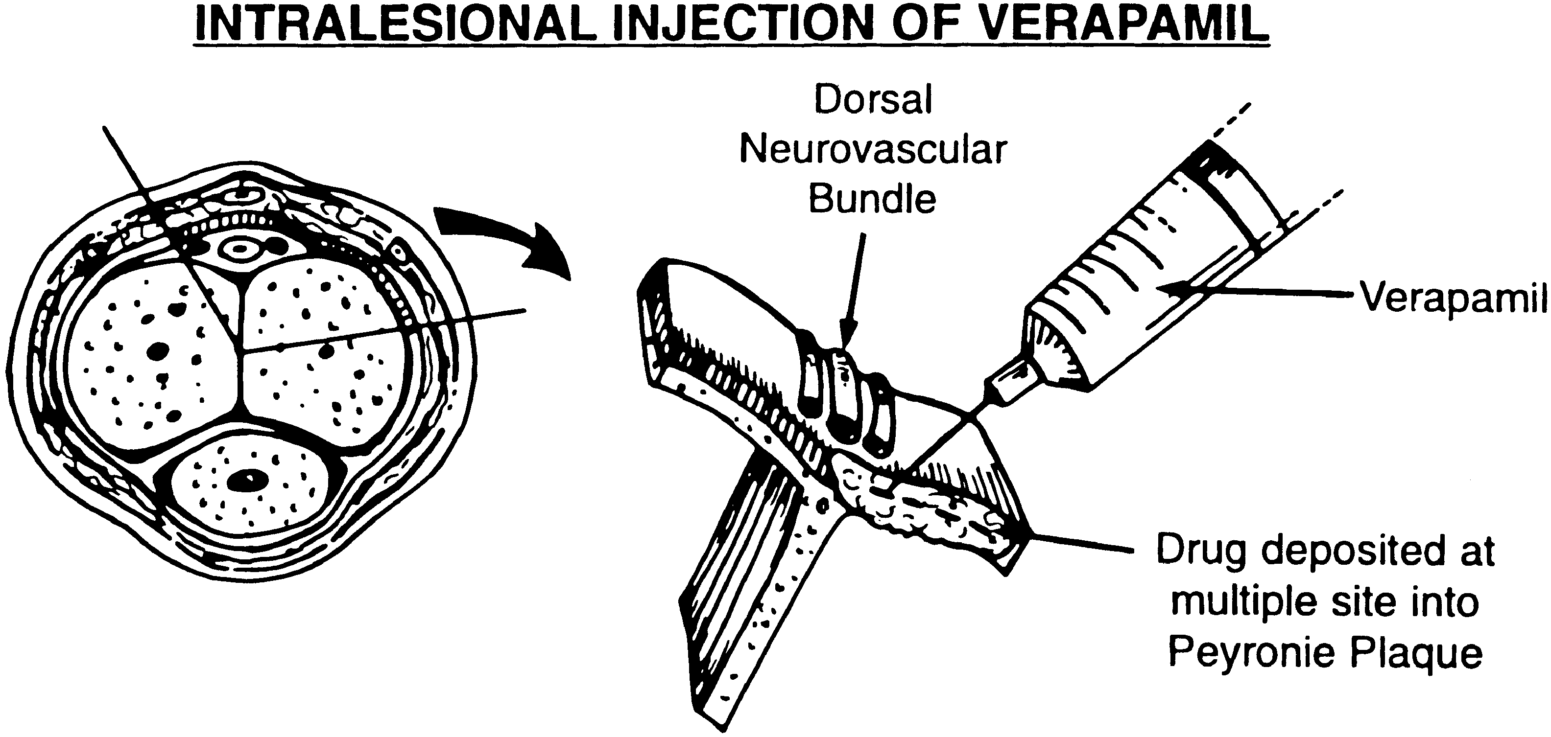 to 6.3 Ϯ 0.44 post-treatment in the control group(P Ͻ0.02).
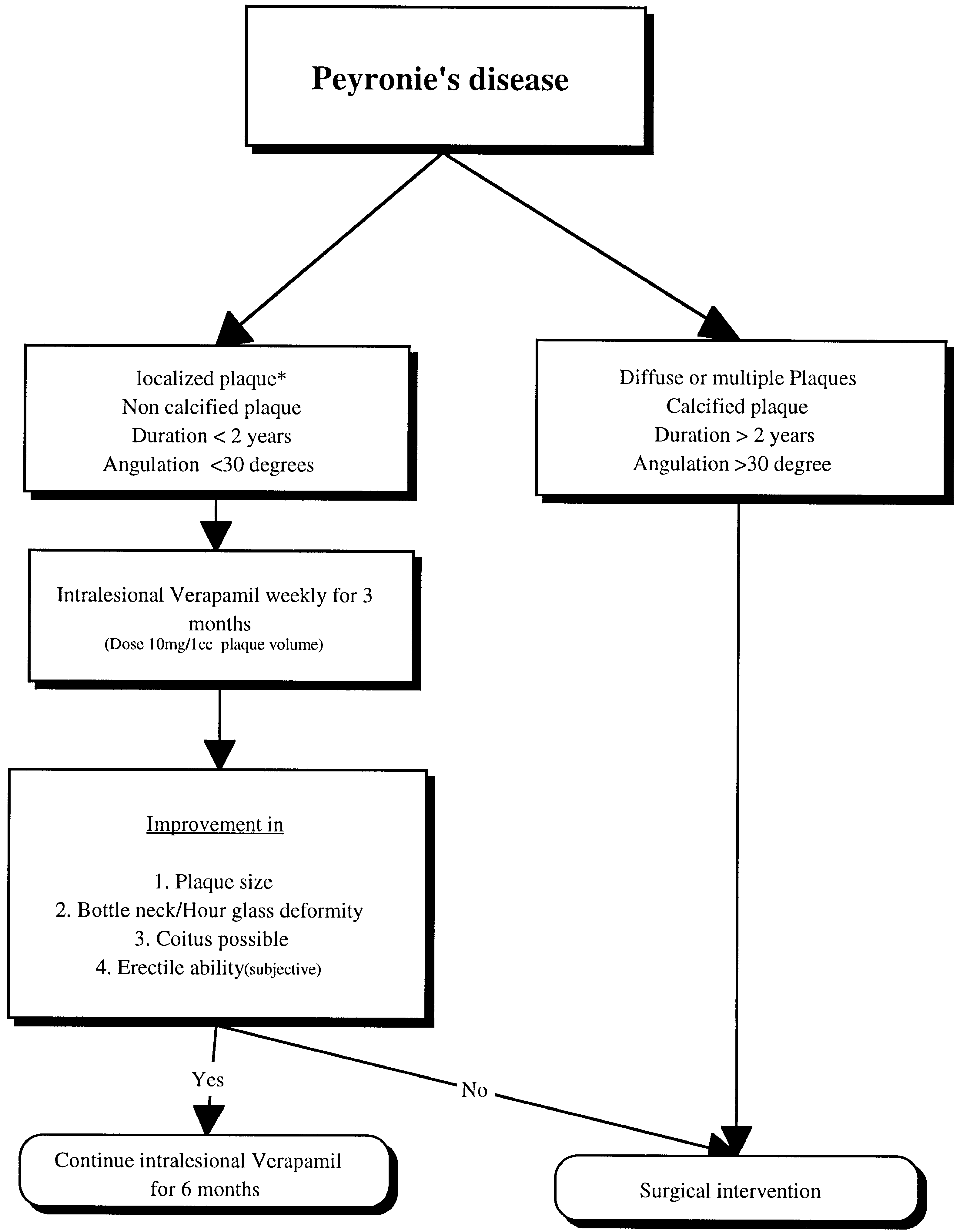 Factors associated in the aetiology of Peyronie’s disease. Br J